
| name | Amanita morenoi |
| name status | nomen acceptum |
| author | Raithelh. |
| english name | "Moreno's Amanita" |
| images |
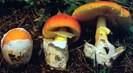
 1. Amanita morenoi, Lago Espejo, Parq. Nac. Nahuel Huapí, Prov. Neuquén, Argentina |
| cap |
Amanita morenoi is a poorly known species. It has a cap 45 - 63 mm wide as dried, pale brown to sordid chestnut brown when fresh, polished, convex when young, and planar in age. The cap margin is apparently nonstriate at first, then short tuberculate-striate. The volva appears on the cap as warts, densest over disc, large, thick, subpyramidal to conical, crowded, brown, and pulverulent. |
| gills |
The gills of this species are free or narrowly adnate, rather close, and cream to white. The short gills are truncate to subtruncate to rounded truncate with attenuate tooth and apparently sparse. |
| stem |
The stem is 68 - 71 × 8 - 10 mm as dried and cream when fresh; and the stem's bulb is 15 - 20 × 14 - 16 mm as dried, possibly subglobose or even subabrupt when fresh; and is hollow as dried in some specimen. There is an membranous, superior, skirt-like annulus that collapses on the stipe. On the stipe the volva is pulverulent brown to chestnut brown and forms scales and warts on lower stipe and the top of the bulb, sometimes warts may be in a ring. |
| odor/taste |
Odor and taste were not recorded for this species as far as we know. |
| spores |
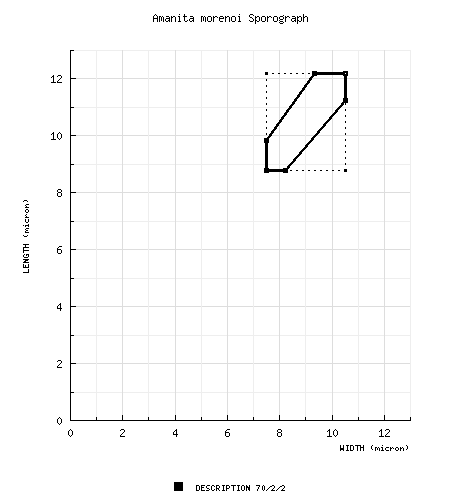
The spores of this species measure (8.4-) 8.8 - 12.2 (-19.6) × (7.0-) 7.5 - 10.5 (-15.4) µm and are subglobose to broadly ellipsoid to ellipsoid (or infrequently globose) and inamyloid. Clamps are rather common at the bases of basidia. |
| discussion |
Amanita morenoi has been found only in Argentina in the area of Lago Espejo in Parque Nacional Nahuel Huapí, in March. If there is a very similar taxon (none are known, the common clamps suggest a possible relationship to the A. muscaria-group), that entity probably also occurs only in the Southern Hemisphere. Specimens of this species have been misdetermined as A. umbrinella E.-J. Gilbert & Cleland, which is a very dark-capped Australian taxon. The species may be toxic and likely to produce symptoms similar to those of A. muscaria (L. : Fr.) Lam. and A. pantherina (DC. : Fr.) Krombh.—R. E. Tulloss |
| brief editors | RET |
| name | Amanita morenoi | ||||||||
| author | Raithelh. 1986. Metrodiana 14: 6, 13 (unnumbered figs.). | ||||||||
| name status | nomen acceptum | ||||||||
| english name | "Moreno's Amanita" | ||||||||
| etymology | genitive of Latinized name, "Moreno's" or "of Moreno" | ||||||||
| MycoBank nos. | 103036 | ||||||||
| GenBank nos. |
Due to delays in data processing at GenBank, some accession numbers may lead to unreleased (pending) pages.
These pages will eventually be made live, so try again later.
| ||||||||
| holotypes | BAFC | ||||||||
| type studies | Tulloss and Halling. 1997. Mycologia 89: 279, figs. 1-8. | ||||||||
| intro |
The following text may make multiple use of each data field. The field may contain magenta text presenting data from a type study and/or revision of other original material cited in the protolog of the present taxon. Macroscopic descriptions in magenta are a combination of data from the protolog and additional observations made on the exiccata during revision of the cited original material. The same field may also contain black text, which is data from a revision of the present taxon (including non-type material and/or material not cited in the protolog). Paragraphs of black text will be labeled if further subdivision of this text is appropriate. Olive text indicates a specimen that has not been thoroughly examined (for example, for microscopic details) and marks other places in the text where data is missing or uncertain. The following description is based on the protolog of the present species, the type study of Tulloss and Halling (1997) and additional, subsequent research by Tulloss. On this tab, the magenta font is used only when specific data is applicable solely to the holotype. | ||||||||
| pileus | 45–63 mm wide as dried, pale brown to sordid chestnut brown, polished, convex when young, planar in age; margin apparently nonstriate at first, then short tuberculate striate; universal veil as warts, densest over disc, large, thick, subpyramidal to conical, crowded, brown, pulverulent. | ||||||||
| lamellae | cream to white, free or narrowly adnate, subclose; lamellulae subtruncate to rounded truncate with attenuate tooth to truncate, apparently sparse (F 1014586). | ||||||||
| stipe | 68–71 × 8–10 mm as dried, cream; bulb 15–20 × 14–16 mm as dried, possibly subglobose or even subabrupt; context hollow as dried in F 1014586; partial veil superior, membranous, skirt-like, collapsing on stipe; universal veil in pulverulent brown to chestnut brown scales and warts on lower stipe and top of bulb, sometimes in ring. | ||||||||
| odor/taste | not recorded. | ||||||||
| macrochemical tests |
KOH - no reaction on context. | ||||||||
| pileipellis | (100–) 140–220 µm thick, colorless at surface, grading to brownish orange toward context; suprapellis 10–50 µm thick, comprising ungelatinized hyphae in gelatinous matrix [with hyphae much denser than in somewhat similar suprapellis of A. flammeola Pegler & Piearce]; subpellis 130–170 µm thick of ungelatinized hyphae; filamentous, undifferentiated hyphae 1.4–12.6 µm wide, gelatinizing, subradially arranged, often constricted at septa, occasionally with slightly thickened walls, occasionally with yellowish subrefractive walls; vascular hyphae (2.0-) 6.5–12.8 µm wide, infrequent or rare. | ||||||||
| pileus context | attacked by mold in holotype; filamentous, undifferentiated hyphae 1.5–11.0 µm wide, plentiful, locally dense, often in fascicles, branching, occasionally with yellowish subrefractive walls; acrophysalides thin-walled, ovoid to subglobose, locally dominating, clavate to ovoid to subglobose, up to 136 × 62 µm; vascular hyphae 2.0–9.8 µm wide, infrequent to locally common. | ||||||||
| lamella trama | bilateral; subhymenial base poorly preserved in holotype, angle of divergence from very shallow to somewhat over 45°, with inflated intercalary cells up to 50 × 21 µm; central stratum not very pronounced at low power magnification, with wcs = 35± µm, consisting largely of chains of inflated hyphal segments up to 38 × 17 µm; filamentous, undifferentiated hyphae 1.8–7.0 µm wide; divergent, terminal inflated cells not observed; vascular hyphae 2.1–11.9 µm wide, locally plentiful, branching. | ||||||||
| subhymenium | wst-near = 15–20 µm; wst-far = 30–50 µm; quite poorly preserved for the most part, with subhymenial tree having branching structure with uninflated and partially inflated hyphal segments dominating (elements at about 45±° to central stratum until rather close to bases of basidia), with inflated cells locally plentiful (thin-walled, up to 26 × 19.0 µm), with basidia arising from subglobose to subpyriform cells arranged in 2 to 3 layers and from partially inflated and uninflated hyphal segments. | ||||||||
| basidia | 38–78 × 7.0–21 µm, [in holotype with plentiful crassobasidia at maturity with walls slightly thickened (up to 0.5 µm thick) and with evenly-spaced hemispherical pits distributed over surface in apical half to third, thin-walled and unpitted when immature, with interior of wall sometimes coated with a layer of refractive yellow material 0.5–0.7 µm thick (sometimes seen in immature basidia without development of thickened wall or pits)], clavate, 2-, 3-, and 4-sterigmate (latter dominant in some regions); clamps relatively common. | ||||||||
| universal veil | On pileus: filamentous, undifferentiated hyphae 1.8–11.2 µm, sometimes with constricted septa, fewer than in warts on stipe base and these concentrated in base of wart and periclinally oriented, frequently branching; inflated cells plentiful to dominating (above wart base), broadly ellipsoid to ellipsoid to subpyriform to clavate to narrowly clavate, up to 84 × 55 µm, occasionally with pale brown contents; vascular hyphae not observed; clamps rare.&nbps; On stipe base: all cells containing yellowish brown pigment; extensively gelatinized or collapsed; filamentous, undifferentiated hyphae 2.0–8.8 µm wide, plentiful, randomly arranged and loosely interwoven; inflated cells ellipsoid to elongate to broadly clavate to cylindric to subfusiform, disordered, terminal, plentiful to locally dominant, up to 67 × 29 µm; refractive hyphae 1.4±–11.0 µm wide, uncommon, fragmented, appearing only on surface (hence probably remains of filamentous, undifferentiated hyphae); clamps present. | ||||||||
| stipe context | longitudinally acrophysalidic; filamentous, undifferentiated hyphae 0.7–9.4 µm wide, occasionally with yellowish subrefractive walls; acrophysalides thin-walled, up to 178 × 49 µm; vascular hyphae 1.4–16.8 µm wide, locally common, sinuous, sometimes curving back on self; clamps present. | ||||||||
| partial veil | known only from holotype; gelatinizing and rather badly damaged by mold [so that we choose not to illustrate it]; filamentous, undifferentiated hyphae 1.0–4.6 µm wide, subradially oriented, dominating; inflated cells scattered, ovoid to broadly clavate, difficult to separate and measure, up to 65? × 25? µm; vascular hyphae 1.4–4.6 µm wide. | ||||||||
| lamella edge tissue | not described. | ||||||||
| basidiospores | [70/2/2] (8.4–) 8.8–12.2 (–19.6) × (7.0–) 7.5–10.5 (–15.4) µm, (L = 10.3–10.5 µm; L’ = 10.4 µm; W = 8.4–9.2 µm; W’ = 8.8 µm; Q = (1.0–) 1.07–1.31 (–1.33); Q = 1.14–1.21; Q’ = 1.18), hyaline, colorless, smooth, thin-walled or with walls up to 0.5 µm thick in normal spores (in crassospores of holotype with wall having 0.8–1.1 µm thick yellow refractive layer appressed to interior of hyaline and slightly thickened spore wall and having latter decorated with small and evenly-spaced hemispherical pits), inamyloid, cyanophilic, globose to subglobose to broadly ellipsoid to ellipsoid, adaxially flattened; apiculus broad and prominent in holotype, relatively small in normal spores, cylindric to narrowly truncate-conic to truncate-conic; contents granular to multiguttulate; color in deposit unrecorded. | ||||||||
| ecology | Known only from around Lago Espejo in Parque Nacional Nahuel Huapí, in March, solitary, growing on ground in ectotrophic forest. Associated vascular plants unrecorded; however, Nothofagus is a possible associate. | ||||||||
| material examined | Tulloss and Halling (1997): ARGENTINA: NEUQUÉN—Parq. Nac. Nahuel Huapí, Lago Espejo, 9.iii.1957 Irma Gamundí s.n. (BAFC 20.572 n.v.; F 1014586 as "A. umbrinella"), 9.iii.1959 Irma Gamundí "Amanita 75)" (holotype, BAFC 30.617). | ||||||||
| discussion |
This taxon was placed in sect. Lepidella by its author. However, this was demonstrated to be in error by Tulloss et al. (1992) and in a type study reported by Tulloss and Halling (1997). The latter authors provide a detailed review of the holotype and of the unusual crassospores and crassobasidia. The portion of the Gamundí collection supposedly deposited in BAFC has not been located. | ||||||||
| citations | —R. E. Tulloss | ||||||||
| editors | RET | ||||||||
Information to support the viewer in reading the content of "technical" tabs can be found here.
Each spore data set is intended to comprise a set of measurements from a single specimen made by a single observer; and explanations prepared for this site talk about specimen-observer pairs associated with each data set. Combining more data into a single data set is non-optimal because it obscures observer differences (which may be valuable for instructional purposes, for example) and may obscure instances in which a single collection inadvertently contains a mixture of taxa.

Text and User-Generated Sporographs are published under the Creative Commons License.

In the case of a taxon page, image credits are on the 'image' tab.